iTero
Our team is proud to utilize the iTero digital dental scanner to create digital impressions of the natural tooth. iTero digital scanner is a revolutionary way to achieve digitized images of the smile for the fabrication of dental restorations and appliances.It offers several benefits, including more accurate representation of the teeth for future procedures, ultimate patient comfort, and the ability to create time-lapse images to show patients the process of certain dental treatments from start to finish. Digital scanners ensure accuracy and allow for easy storage and access of image files stored at our office.

CBCT
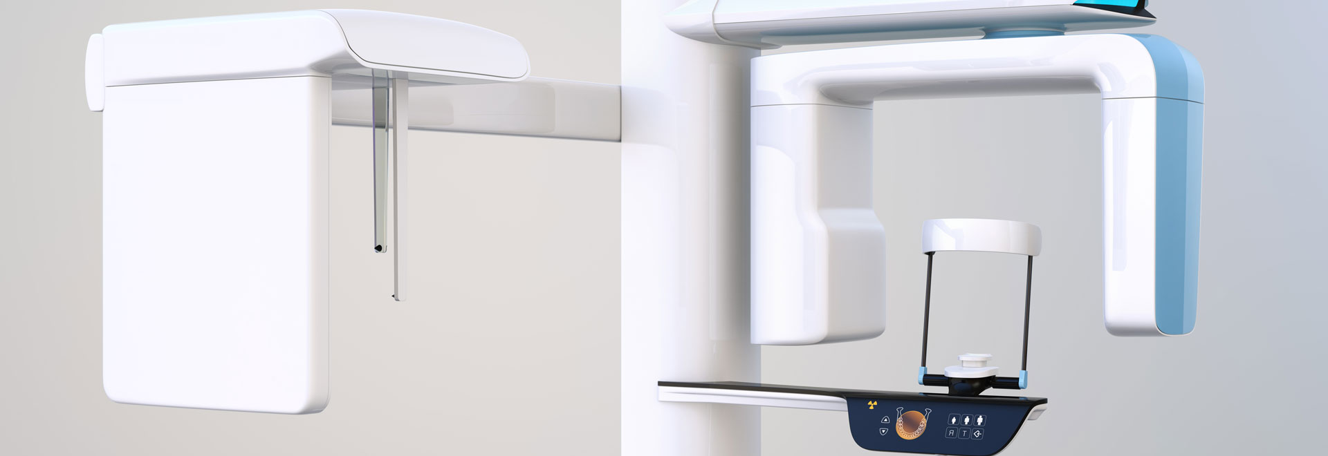
Cone-beam computed tomography systems are radiographic systems used by dental professionals to analyze and reconstruct 3D images of a patient’s teeth, jaws and surrounding anatomy. The information obtained by means of CBCT imaging is useful in both diagnosis and precise treatment planning when two-dimensional diagnostic films are insufficient. Dental CBCT is useful for multiple types of analyses as well as the assessment of maxillofacial disorders or pathology. It is also most useful in surgical planning, including the accurate placement of dental implants.
Our office utilizes the Planmeca Promax Ultra Low Dose® machine. This is the leading method for acquiring CBCT images at a low patient dose.

Digital Radiography
Digital radiography utilizes computer technology and digital sensors for the acquisition, viewing, storage, and sharing of radiographic images. It offers several advantages over the older traditional film based methods of taking x-rays. The most significant of these advantages is that digital radiography reduces a patient’s exposure to radiation. Other benefits are that images can be viewed instantly after being taken, can be seen simultaneously as needed by multiple practitioners, and can be easily shared with other offices. Digital x-rays are also safer for the environment as they do not require any chemicals or paper to develop.
An electronic pad, known as a sensor is used instead of film to acquire a digital image. After the image is taken, it goes directly into the patient’s file on the computer. Once it is stored on the computer, it can be easily viewed on a screen, shared, or printed out.

Diagnodent
Using the Diagnodent instrument, our team can easily detect cavities in teeth, even in their earliest stages. This device allows us to find areas of decay sooner for more effective intervention, treating cavities before they become a bigger, more damaging problem for oral health and wellness. While patients relax in the treatment chair, they will be able to have their smile evaluated for cavities with the Diagnodent device, which reflects a different color of light back when a tooth has a cavity. X-rays can then be used to pinpoint the cavity in the mouth and determine what treatment is best. With the Diagnodent, we can provide earlier diagnosis of conditions to reduce the risk of extensive damage to the smile by placing fillings when necessary.


